Lens Anatomy
Lens shape and structure
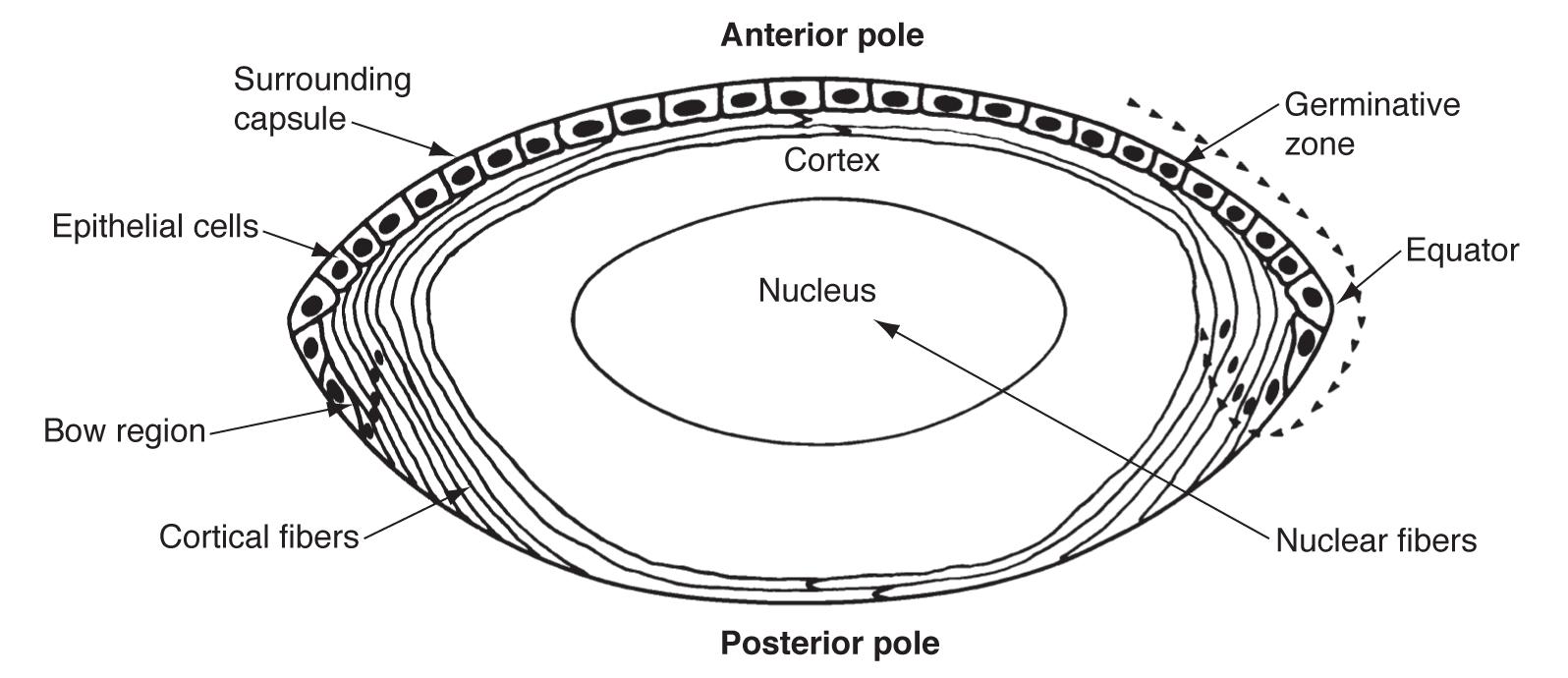
The lens is a biconvex, avascular, noninnervated, encapsulated body composed entirely of epithelial cells and fibers. The 3 layers of the lens are the nucleus, cortex, and capsule. Lens transparency depends on tight, regular packing of lens fibers and their intracellular proteins to permit light transmission and provide refractive power for accommodation (focusing on near objects).
Anatomical position of the lens
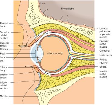
The lens is suspended just posteriorly to the iris and divides the anterior and posterior segments of the eye. The anterior segment is the first third of the eye from the cornea to the lens. The anterior segment includes the iris, ciliary body, and two fluid filled chambers, also called anterior and posterior. The anterior chamber includes the space between the iris and the endothelial layer of the cornea, while the posterior chamber includes the space between the posterior iris, zonules, and posterior lens. Both chambers are filled with aqueous humor, which is produced by the ciliary epithelium and flows over the lens around the iris to be reabsorbed by the trabecular meshwork.
The posterior segment is the posterior two-thirds of the eye, from the hyaloid membrane just posterior to the lens and spanning all the way to the optic nerve. This includes the vitreous humor, retina, choroid, and optic nerve.
The posterior surface of the lens itself is attached to the vitreous humor at Wieger’s Ligament (also known as Hyaloideo capsulare). The potential space between the lens capsule and hyaloid face of the vitreous is the retrolental space (or Burger’s space).
Lens zonules
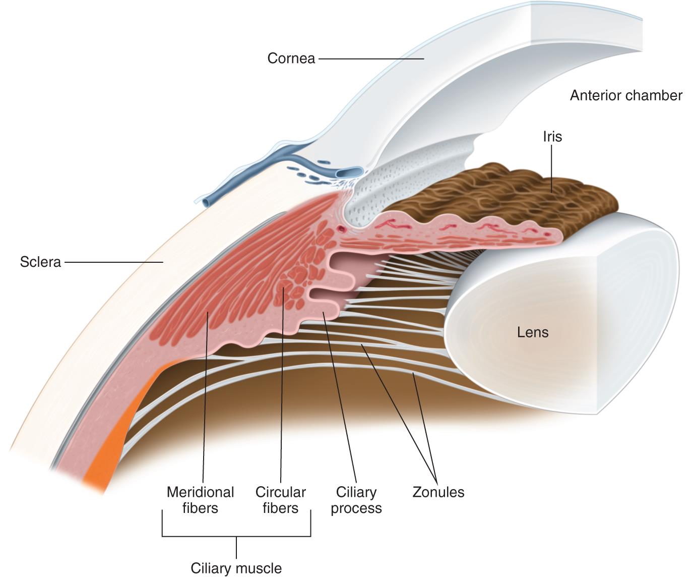 Zonular fibers attach to the outer layer of the lens capsule (the zonular lamella) and hold the lens in place. Zonular fibers originate in the basal lamina of the pars plana and pars plicata of the ciliary body and insert on the equatorial region of the lens.